|
Hi Visitor
In this and the next eZine, we are exploring the clinical usefulness of musculoskeletal ultrasound or for those into acronyms, MSK US. These case studies have been kindly provided by Mr Peter Esselbach course presenter of our MSK US course. Move your scroll icon over each picture for associated text.
Case Study 1

Sean is a 52 year old male who reported pain in his right upper arm after being pulled from the water abruptly while water skiing. He noticed bruising develop in his right cubital fossa and experienced pain with putting his right arm into internal rotation. He also had pain abducting his right arm above 90 degrees. His GP sent him for investigations and treatment, thinking he had a tear to his distal biceps brachii muscle.
I examined him 1 week after the accident. He had no palpation tenderness in his cubital fossa and diagnostic ultrasound revealed a normal distal biceps brachii tendon.
He had tenderness of the right upper arm along the right axillary line. Ultrasound images of the upper right arm are shown (see Figure 2 & 3) 
The pectoralis major fibres were followed on ultrasound to reveal a normal pectoralis major tendon. The diagnosis was then made of a Grade II tear of the musculotendinosis junction of the right sternal head of the pectoralis major. An ultrasound of the subdeltoid region revealed a normal rotator cuff but a thickened subacromial-subdeltoid bursa (see Figure 4).
 In addition to the pectoral tear, he had subacromial-subdeltoid bursitis. He was treated with antiinflammatory medication for 5 days to help settle his pain. Given the ultrasound revealed no tendinous tear, no surgical intervention was required. Instead he was advised to rest , avoiding any activity provoking pain. He was gradually progressed onto a stretching and strengthening rehabilitation program. In addition to the pectoral tear, he had subacromial-subdeltoid bursitis. He was treated with antiinflammatory medication for 5 days to help settle his pain. Given the ultrasound revealed no tendinous tear, no surgical intervention was required. Instead he was advised to rest , avoiding any activity provoking pain. He was gradually progressed onto a stretching and strengthening rehabilitation program.
Case Study 2

Anne is a 70 yo lady with right posterior heel pain and a history of insertional calcific tendinopathy. She had previous treatment to the heel with ultrasound guided cortisone which had no effect and shockwave therapy which after 6 weeks had a 90 % reduction in her pain. She presented 7 months later reporting her pain had returned in the same area.
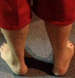
On observation see had swelling of her right posterior heel (see Figure 1), a reduced stride length and limited dorsiflexion with push-off phase in her gait cycle.
An ultrasound revealed a swollen right Achilles tendon with areas of partial tears and calcific tendinopathy at the insertion (see Figure 2) . Colour doppler also demonstrated increased vascularity in the tendon indicating the chronicity of the pathology (see Figure 3).
 
Further evaluation of Anneʼs lower limb showed marked reduction in her medial gastrocnemius muscle bulk and fatty infiltration suggesting a lack of activation of the muscle during the gait cycle.
.
 Physiotherapy treatments involved dry needling the calf and heel to reduce pain as well as soft tissue techniques to decrease myofascial tone in the calf. Advice was given as to appropriate foot wear and a heel raise was added into her right shoe. A home exercise program of double leg (for 6 weeks) and then single leg (for 10 weeks) isometric heel raises on the flat ground was given. Anne reported a 80% reduction in her pain scores and had increased her walking tolerance to 2 km. Physiotherapy treatments involved dry needling the calf and heel to reduce pain as well as soft tissue techniques to decrease myofascial tone in the calf. Advice was given as to appropriate foot wear and a heel raise was added into her right shoe. A home exercise program of double leg (for 6 weeks) and then single leg (for 10 weeks) isometric heel raises on the flat ground was given. Anne reported a 80% reduction in her pain scores and had increased her walking tolerance to 2 km.
I was fortunate to be able to attend Peter's course last year and quickly appreciated the unique skill he has to offer because of his training in sonography and physiotherapy. If you are interested in developing your MSK US skill, then I doubt you will find a better opportunity in Australia. Click RT and MSK US Course for course details.
Recent Blogs of Interest
All the best,
Doug Cary FACP APAM
Specialist Musculoskeletal Physiotherapist
You are free to use material from the Clinical Kit eZine in whole or in part, as long as you include complete attribution, including live website link. Please also notify me where the material will appear. The attribution should read: "By Doug Cary FACP of AAP Education. Please visit our website at www.aapeducation.com.au for additional clinical articles and resources on post graduate education for health professionals" (Please make sure the link is live if placed in an eZine or in a web site.)
If you would like to unsubscribe click {unsubscribe}Unsubscribe{/unsubscribe}
HOME | DRY NEEDLING | INTEGRATED NECK SOLUTIONS | 100% GUARANTEE | MULLIGANS | BLOG | MSK US
Like {share:facebook} Share {share:twitter}
|

